热线电话 Hot-line

18717985772

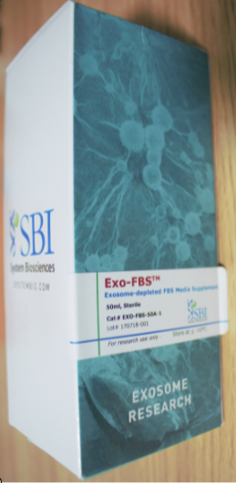
Bovine exosome-depleted FBS, for isolating exosomes from cultured cells without the complication of bovine exosomes that are present in conventional FBS

品牌SBI
货号EXO-FBS-50A-1/EXO-FBS-250A-1
英文名称Bovine exosome-depleted FBS
规格
  • 50ml
  • 250ml
  • 价格
    • 2300
    • 询价

    Overview

    Avoid inadvertently studying bovine exosomes

    Fetal bovine serum, or FBS, is an important component of many types of cell culture media. But for researchers interested in isolating exosomes from cultured cells, standard FBS can introduce unwanted complications—bovine exosomes, which can cause significant background issues or interfere with functional studies. Which is why SBI developed Exo-FBS, our patented exosome-depleted FBS.

    • Exosome-sized vesicles removed
    • Very low levels of CD63-positive bovine exosomes
    • Undetectable levels of bovine miRNAs
    • Comparable growth rates as standard FBS
    • Interchangeable with standard FBS (add 10% in DMEM or RPMI)


    Supporting Data

    High quality and great performance

    Exo-FBS supports robust cell growth (Figure 1), has greatly reduced levels of bovine exosomes (Figures 2), and bovine miRNAs (Figure 3). Cell growth in media supplemented with Exo-FBS is similar to cell growth in media supplemented with standard FBS (Figure 1).

    Exo-FBS supports more robust cell growth health than competitors

    Exo-FBS supports more robust cell growth than competitors

    Figure 1. Exo-FBS supports more robust cell growth than Company G’s exosome-depleted FBS. HepG2 cells were grown in media containing either standard FBS (control), Exo-FBS, or Company G’s exosome-depleted FBS. Data is shown after one and two passages and illustrates that Exo-FBS provides a higher number of viable cells (left panel, viability assessed using a CCK-8 assay) and a healthier cell morphology (right panel) than Company G’s product.


    anti-CD63 Elisa shows low levels of exosomes in Exo-FBS


    Figure 2. Bovine α-CD63 ELISA shows low levels of exosomes in Exo-FBS CD63 is an exosome-specific marker. An α-CD63 ELISA of standard FBS and Exo-FBS shows very low levels of CD63 in Exo-FBS, supporting the NTA data, which showed low numbers of exosome-sized particles in Exo-FBS (Figure 1). Equal volumes (50 µl) of either standard FBS or Exo-FBS depleted media supplement were used and the graphed results normalized to the signal level of standard FBS.


    qPCR shows bovine miRNA is undetectable in Exo-FBS


    Figure 3. qPCR assays show undetectable levels of bovine exosomal miRNAs in Exo-FBS. While standard FBS contains amplifiable miRNAs (12 of the 72 individual miRNAs tested, left panels), Exo-FBS shows no amplifiable miRNAs (right panels). Standard FBS and Exo-FBS media supplements (4 ml) were treated with Trizol extraction methods to recover exosomal RNAs. RNA was converted to cDNA and 72 individual bovine microRNAs were measured by qPCR using SBI’s QuantiMir system.



    Product Documentation

    Citations

    • Akerman, AW, et al. (2019) Elevated Wall Tension Leads to Reduced miR-133a in the Thoracic Aorta by Exosome Release. J Am Heart Assoc. 2019 Jan 8; 8(1):e010332. PM ID: 30572760
    • Raffo-Romero, A, et al. (2018) Medicinal Leech CNS as a Model for Exosome Studies in the Crosstalk between Microglia and Neurons. Int J Mol Sci. 2018 Dec 19; 19(12). PM ID: 30572617
    • Guo, D, et al. (2018) RAB27A promotes melanoma cell invasion and metastasis via regulation of pro-invasive exosomes. Int. J. Cancer. 2018 Dec 17;. PM ID: 30556600
    • Busatto, S, et al. (2018) Tangential Flow Filtration for Highly Efficient Concentration of Extracellular Vesicles from Large Volumes of Fluid. Cells. 2018 Dec 16; 7(12). PM ID: 30558352
    • Almanza, G, et al. (2018) Extracellular vesicles produced in B cells deliver tumor suppressor miR-335 to breast cancer cells disrupting oncogenic programming in vitro and in vivo. Sci Rep. 2018 Dec 4; 8(1):17581. PM ID: 30514916
    • Lainšček, D, et al. (2018) Delivery of an artificial transcription regulator dCas9-VPR by extracellular vesicles for therapeutic gene activation. ACS Synth Biol. 2018 Dec 4;. PM ID: 30513193
    • Lee, H, et al. (2018) pH-responsive hyaluronate-anchored extracellular vesicles to promote tumor-targeted drug delivery. Carbohydrate Polymers. 2018 Dec 1; 202:323-333. Link: Carbohydrate Polymers
    • Shtam, T, et al. (2018) Plasma exosomes stimulate breast cancer metastasis through surface interactions and activation of FAK signaling. Breast Cancer Res Treat. 2018 Nov 27;. Link: Breast Cancer Res Treat
    • Kanlikilicer, P, et al. (2018) Exosomal miRNA confers chemo resistance via targeting Cav1/p-gp/M2-type macrophage axis in ovarian cancer. EBioMedicine. 2018 Nov 25;. PM ID: 30487062
    • Peng, Q, Zhang, J & Zhou, G. (2018) Circulating exosomes regulate T-cell mediated inflammatory response in oral lichen planus. J Oral Pathol Med. 2018 Nov 17;. Link: J Oral Pathol Med
    • Peng, Q, Zhang, J & Zhou, G. (2018) Circulating exosomes regulate T-cell-mediated inflammatory response in oral lichen planus. J. Oral Pathol. Med.. 2018 Nov 17;. PM ID: 30447107
    • Mrowczynski, OD, et al. (2018) Exosomes impact survival to radiation exposure in cell line models of nervous system cancer. Oncotarget. 2018 Nov 16; 9(90):36083-36101. PM ID: 30546829
    • Li, B, et al. (2018) Effects of tumor necrosis factor-α-induced exosomes on the endothelial cellular behavior, metabolism and bioenergetics. Microcirculation. 2018 Nov 15;:e12515. PM ID: 30431204
    • Sun, H, et al. (2018) Macrophages alternatively activated by endometriosis-exosomes contribute to the development of lesions in mice. Mol. Hum. Reprod.. 2018 Nov 14;. PM ID: 30428082
    • Lehrich, BM, et al. (2018) Fetal Bovine Serum-Derived Extracellular Vesicles Persist within Vesicle-Depleted Culture Media. Int J Mol Sci. 2018 Nov 9; 19(11). PM ID: 30423996
    • Tang, YT, et al. (2018) Alterations in exosomal miRNA profile upon epithelial-mesenchymal transition in human lung cancer cell lines. BMC Genomics. 2018 Nov 6; 19(1):802. PM ID: 30400814
    • Hu, J, et al. (2018) Exosomal Mst1 transfer from cardiac microvascular endothelial cells to cardiomyocytes deteriorates diabetic cardiomyopathy. Biochimica et Biophysica Acta (BBA) - Molecular Basis of Disease. 2018 Nov 1; 1864(11):3639-3649. Link: Biochimica et Biophysica Acta (BBA) - Molecular Basis of Disease
    • Hu, W, et al. (2018) Adipose tissue browning in cancer-associated cachexia can be attenuated by inhibition of exosome generation. Biochem. Biophys. Res. Commun.. 2018 Oct 16;. PM ID: 30340833
    • Mao, Q, et al. (2018) GMSC-Extracellular Vesicles Activate Schwann Cell Repair Phenotype and Promote Nerve Regeneration. Tissue Eng Part A. 2018 Oct 12;. PM ID: 30311853
    • Sun, H, et al. (2018) Eutopic stromal cells of endometriosis promote neuroangiogenesis via exosome pathway. Biol. Reprod.. 2018 Oct 5;. PM ID:30295741
    点击关闭
    • 觅拓-达斐

      QQ

      公众号